Cumhuriyet Mh. Büşra Sk. No: 18/20 (Ozan Plaza) Süleymanpaşa/TEKİRDAĞ


Panoramic, Cephalometric, and 3D Tomography Imaging
Panoramic, cephalometric, and three-dimensional tomography imaging methods are advanced radiological diagnostic techniques that enable detailed evaluation of the teeth, jaws, and surrounding tissues. These imaging techniques are of great importance for accurate diagnosis and effective treatment planning. They can be applied quickly and safely in a clinical setting.
What is panoramic imaging?
Panoramic imaging is a radiological method that allows all teeth, jaw bones, and temporomandibular joints to be evaluated on a single image. It provides general information about impacted teeth, cavities, cysts, and bone structures. It is frequently used in routine pre-treatment evaluations.
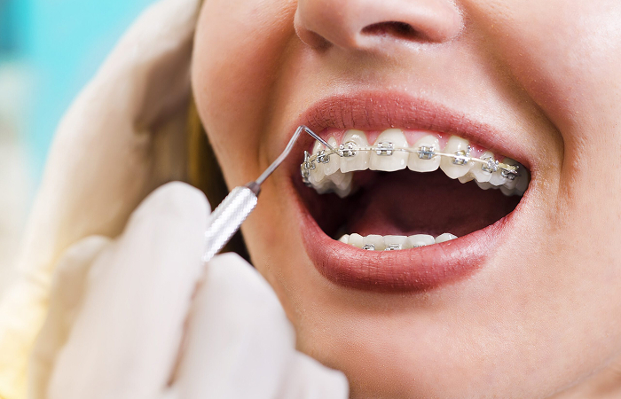
In which cases is cephalometric imaging used?
Cephalometric imaging is a method used especially in orthodontic treatment planning. It enables the analysis of jaw relationships, tooth positions, and facial skeletal structure. It allows for comparative evaluation before and after treatment.
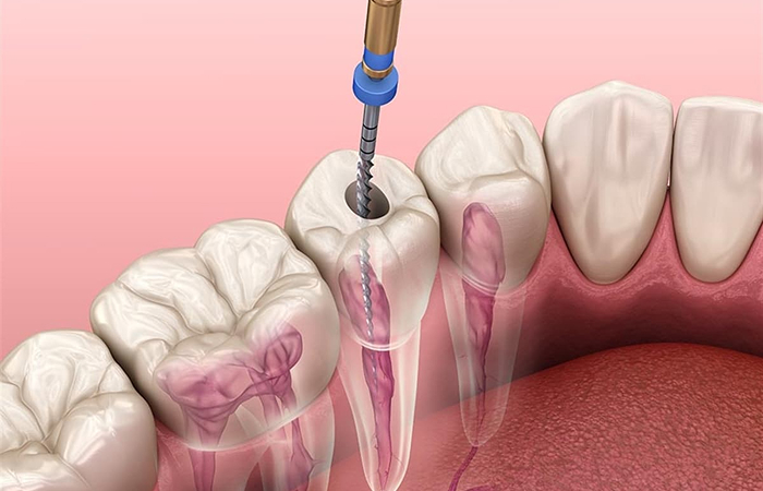
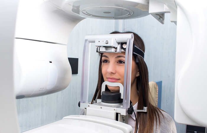
In which cases is 3D tomography imaging preferred?
Three-dimensional tomography is preferred for implant planning, impacted tooth surgery, detailed examination of bone structures, and advanced surgical procedures. Sinuses, nerve canals, and bone density can be clearly visualized. This helps minimize surgical risks.
Things to Consider Before and After Imaging
Metal accessories should be removed before imaging. Pregnancy status must be reported to the physician. There are no restrictions after the procedure. The obtained images are safely used in treatment planning and enable accurate and effective interventions.